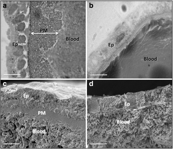
Fig. 1.

Morphology of An. aquasalis midguts after normal and chitinase-containing blood meals. Histology (a) and SEM (c) of An. aquasalis midguts 24 h after a normal blood meal. The thick PM is visible isolating the midgut epithelium from the partially digested blood meals. Similar histology (b) and SEM (d) of An. aquasalis midguts 24 h after a chitinase-containing blood meal. The PM is absent inside the midgut, and the blood meal is in direct contact with the epithelium. Abbreviations: PM, peritrophic matrix; Ep, epithelium; Blood, blood meal. Scale-bars: a-d, 50 μm
