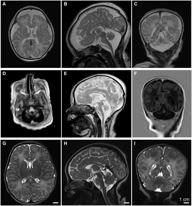
Figure 1.
Neuroimaging features in two index patients with PCH. (A) Axial, (B) sagittal, and (C) coronal T2-weighted images from the index patient from Family 1 performed on Day 5 of life, demonstrating small cerebellum, pons, and medulla, with prominent ventricles and increased extra-axial spaces, and a thin corpus callosum. (D) Axial, (E) sagittal, and (F) coronal images from the similarly affected index patient from Family 2, performed on Day 6 of life, demonstrating pontocerebellar hypoplasia, prominent ventricles and increased extra-axial spaces, a thin corpus callosum, compared to normal findings from (G) axial, (H) sagittal, and (I) coronal T2-weighted images from a normal subject at 8 days of age.

