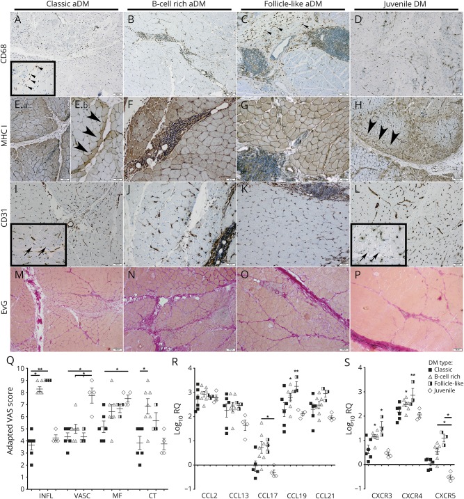
Figure 2. Histopathologic evaluation of sections of muscle biopsy specimens based on 4 different domains.
Muscle specimens were evaluated based on 4 domains (inflammatory [INFL], vascular [VASC], muscle fiber [MF], and connective tissue [CT]). In jDM and classic aDM, CD68+ macrophages were diffusely distributed in the perimysium and adjacent endomysium (A, arrowheads; D). In B-cell–rich and follicle-like aDM, CD68+ macrophages were predominantly seen in the periphery of the lymphocytic aggregates (B, C, arrowheads). Expression of MHC class I molecules was predominantly seen in the perifascicular region of jDM and classic aDM muscle samples (2 patients: Ea and Eb, arrowheads; H, arrowheads) and in B-cell–rich and follicle-like aDM on the sarcolemma of all muscle fibers with perifascicular enhancement (F, G). CD31 highlighted the capillary loss, which was most prominent in the perifascicular region in jDM (L, arrows) and milder in classic (I, arrows), B-cell–rich, and follicle-like aDM (J, K). Elastica van Gieson (EvG) staining was used to evaluate IM fibrosis (M–P), which was more prominent in B-cell–rich (N) and follicle-like aDM (O) than in jDM (P) and classic aDM (M). (Q) Results of the evaluation of the adapted VAS score (higher scores = more disease activity). (R, S) mRNA expression of chemokines CCL2,-13,-17,-19,-21 and chemokine receptors CXCR3,-4,-5 in different DM subgroups was measured by real-time PCR (RT-PCR). The ΔCT of healthy controls was subtracted from the ΔCT of patients with DM to determine the differences (ΔΔCT) and fold change (2^−ΔΔCT) of gene expression. Gene expression was illustrated by the log10 of fold change values compared with the normal controls. Results show mean ± SEM. *p < 0.05, **p < 0.01. aDM = adult dermatomyositis; DM = dermatomyositis; IM = intramuscular; jDM = juvenile dermatomyositis; mRNA = messenger RNA.

