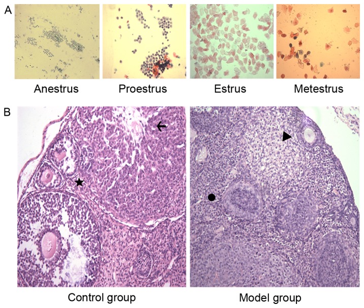
Figure 1.

(A) Normal mouse vaginal smears (magnification, ×100; pap staining; yellow filter). (B) Ovarian pathology (magnification, ×200; hematoxylin and eosin staining) prior to and following modeling compared with the control group. The images revealed that there were primordial follicles, and fewer typical growing and mature follicles in the cortex of mouse ovaries in the model group compared with the control group, and a large number of apoptotic cells were observed in these follicles. Primary follicles are indicated by a star; corpus luteum is indicated by an arrow; primordial follicle is indicated by a triangle; and a dysplastic follicle is indicated by the circle.
