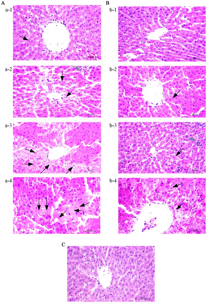
Figure 2.
Thymosin α1 treatment attenuates hepatic tissue injury in D-galactosamine hydrochloride/lipopolysaccharide-induced ALF. Liver tissues were harvested at 3, 6, 9 and 12 h after the establishment of ALF for histopathological examination (magnification, ×400). Representative images from 3 rats/group were selected. (A) MG at A-1, 3 h; A-2, 6 h; A-3, 9 h; and A-4, 12 h. (B) TG at B-1, 3 h; B-2, 6 h; B-3, 9 h; and B-4, 12 h. (C) CG 3 h. Black arrows indicate apoptosis and necrosis, grey arrows indicate inflammatory cell infiltration. ALF, acute liver failure; CG, control group; MG, model group; TG, treatment group.

