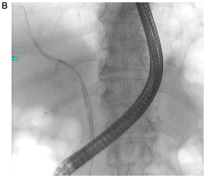
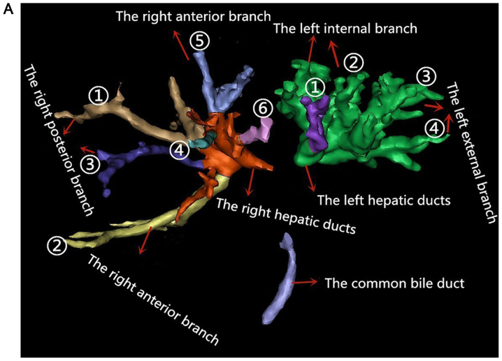
Figure 4.

Bile ducts in 3DV model and the image of ERCP. (A) The liver bile duct system without the tumor and the bile ducts identified by different colors. In the right bile ducts, 1 and 3 belong to the right posterior branch, and 2 and 4–6 belong to the right anterior branch. In the left bile ducts, 1 and 2 belong to the left internal branch, and 3–5 belong to the left external branch. (B) The guide wire was inserted into the right hepatic duct in ERCP operation. 3DV, three-dimensional visualization; ERCP, endoscopic retrograde cholangiopancreatography.

