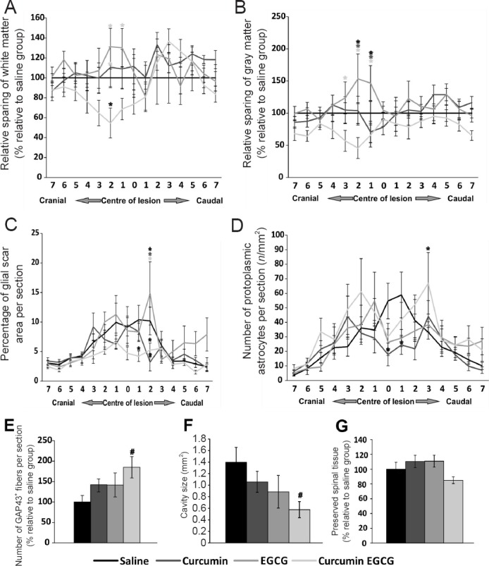
Figure 2.

Histological and immunohistochemical analysis results of injured spinal cords.
The amount of spared white (A) and grey matter (B) was determined 9 weeks after spinal cord injury (SCI) in rats treated with saline, curcumin, EGCG, or their combination. No statistically significant effect of applied drugs on tissue preservation was observed. In addition, the volume of white matter and gray matter across the measured regions revealed no significant difference (G). However, a strong trend toward smaller cavity size was found (F). The effect of the application of curcumin, EGCG, or their combination on glial scar formation (C), and the average number of protoplasmic astrocytes per section (D) around the central lesion cavity was measured 9 weeks after SCI. The combined application of curcumin and EGCG had a suppressive effect on glial scar formation at the area of the lesion epicenter. In the central region of the injury, all treatments showed a positive effect on decreasing the number of protoplasmic astrocytes. The distribution of the effect on X axis is measured as distance in mm from the lesion center (set as 0) (A–D). The effect of applied curcumin, EGCG, or their combination on axonal sprouting. (E) The combination of curcumin and EGCG had a synergistic effect on axonal sprouting, when compared to saline treated animals. Two-way repeated measures analysis of variance (ANOVA) with Student-Newman-Keuls test was used to determine statistical significance (A–D). One-way ANOVA with Student-Newman-Keuls test was used to determine the level of statistical significance (E–G). *P < 0.05 (the color of asterisk indicates the statistically different group(s)). Symbol of # is used when trend toward significance has reached statistical value P = 0.05–0.06. n = 5 per group.
