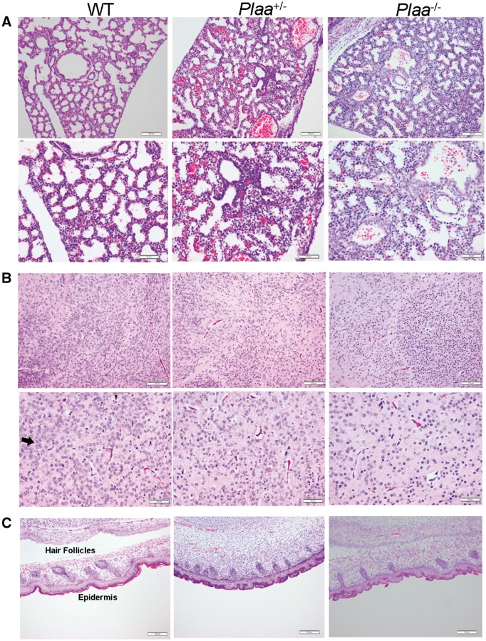
Figure 6.
Histopathology of embryonic mouse tissues (5 µm) at embryonic Day 18.5. Lungs (A), brain cerebral cortex (B), and skin (C) were haematoxylin and eosin stained and analysed in a blinded fashion. Tissues representing two wild-type, two Plaa+/−, and four Plaa−/− embryos were analysed. Multiple fields for each tissue were visualized and typical representations are shown with scale bars of 100 µm (magnification ×100; top rows in A–C) and 50 µm (maginification ×200; bottom rows in A and B). Arrows in the haematoxylin and eosin stained slides indicate examples of mature neurons, while the arrowheads indicate dark round cells as examples of immature neurons or oligodendroglia. Plaa−/− mouse embryos showed an increasing number of less matured and undifferentaited neurons.

