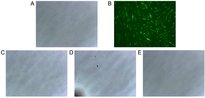
Figure 1.
Overexpression of Sema3A is successfully achieved by vector infection and the cell morphology is examined. Representative overlay images of (A) phase contrast and (B) fluorescence microscopy demonstrated the transduction efficiency of Sema3A for green fluorescent protein using adenoviral transduction. The cell morphology of the (C) untreated control cells, (D) cells infected with pCMV-MCS-EGFP (control vector) and (E) cells infected with pAdCMV-SEMA3A-MCS-EGFP (Sema3A overexpressing vector) was examined under an electron microscope. Experiments were performed in triplicate. Sema3A, semaphorin 3A.

