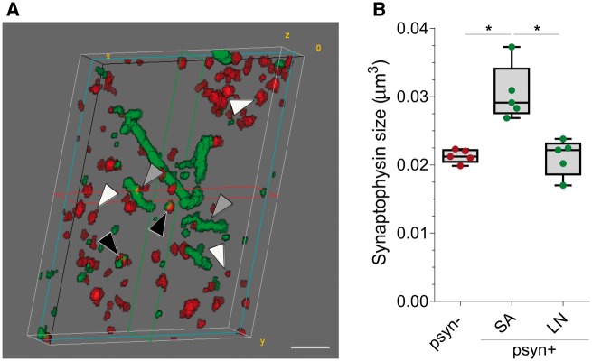
Figure 3.
Median volume of presynaptic terminals co-localizing with p-α-synuclein. (A) 3D reconstruction of 20 consecutive sections from a DLB case. P-α-synuclein (green) and synaptophysin (red) are shown. Black arrowheads indicate synaptophysin objects that co-localize with small aggregates of p-α-synuclein, grey arrowheads point to synaptic terminals that co-localize with Lewy neurites, and white arrowheads those synaptophysin objects that do not co-localize with p-α-synuclein. (B) Median volumes of synaptic terminals according to co-localization with p-α-synuclein (p-α-synuclein +, or −) and their characteristic pattern (small aggregate or Lewy neurite, *P < 0.05). LN = Lewy neurite; Psyn = p-α-synuclein; SA = small aggregate. Scale bar in A = 2 µm.

