Figure 4.
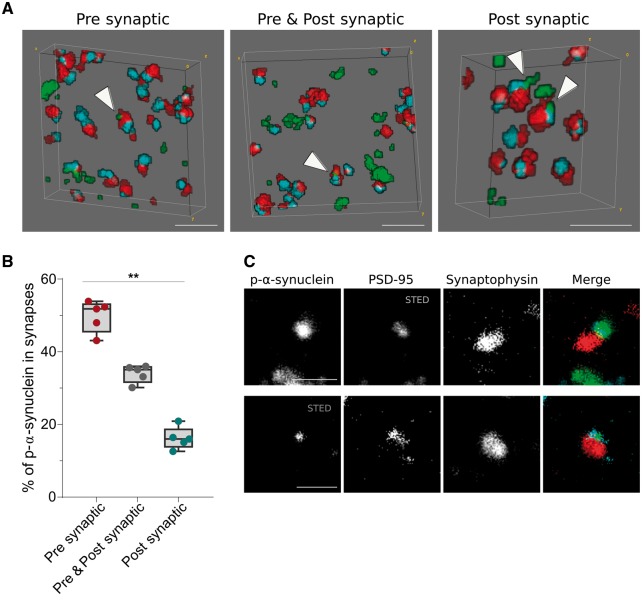
Trans-synaptic localization of p-α-synuclein. (A) 3D reconstruction of 20 consecutive sections from a DLB case. P-α-synuclein (green), synaptophysin (red) and PSD-95 (blue) are shown. White arrowheads point to zones where p-α-synuclein co-localizes with synaptophysin (presynaptic), PSD-95 (postsynaptic) or both (pre- and postsynaptic). In B, p-α-synuclein objects that co-localized with synaptic pairs (synaptophysin and PSD-95 objects) are classified depending on its presynaptic, pre- and postsynaptic or postsynaptic localization. The relative co-localization of p-α-synuclein with the synaptophysin presynaptic marker was significantly higher than the PSD-95 postsynaptic co-localization. (C) Representative images of the co-localization between between p-α-synuclein, synaptophysin and PSD-95 using array tomography combined with STED microscopy. A single 70 nm-thick section stained for p-α-synuclein (green), synaptophysin (red) and PSD-95 (blue) is shown. STED was applied to image PSD-95 (top row) or p-α-synuclein (bottom row). Scale bar in A = 2 µm; C = 1 µm.

