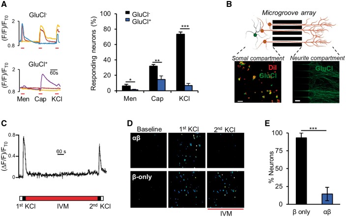
Figure 2.
GluCl mediates local silencing in response to excitatory stimuli. (A, left) Representative somal Ca2+ responses of ivermectin-treated DRG neurons to consecutive stimulation with 200 μM menthol (Men), 1 μM capsaicin (Cap) and 50 mM KCl. (Right) Proportion of neurons that responded to each stimuli. One-way ANOVA with post hoc Bonferroni test. Data derived from n = 9 independent experiments (GluCl+, total of 162 neurons, GluCl−, 629 neurons). (B) Schematic of experimental set-up illustrating somal and neurite compartments separated by a microgroove array. DiI treatment of the neurite compartment marks GluCl+ cell bodies that have successfully sent axonal projections through the microgroove array. GluCl+ axons are clearly discernable in the neurite compartment. Scale bars = 50 µm. (C) Somal Ca2+ transients of a control neuron in response to the experimental stimulation protocol [two KCl (50 mM) neurite stimulations separated by 12 min ivermectin (20 nM) wash-on only to the neurite compartment]. This set-up allows us to assess the effect of local drug administration to axonal activation. (D) Ratiometric Ca2+ responses of DRG cell bodies while neurites are stimulated in isolated compartments, before and after acute ivermectin treatment of neurites. (E) Percentage of cell bodies that responded to the second neurite depolarization stimulus, as a proportion of those that responded to the first stimulation. Student’s unpaired t-test, n = 4 independent experiments (β-only, total of 138 cells, αβ, 200 cells). All grouped data are mean ± SEM, *P < 0.05, **P < 0.01, ***P < 0.001.

