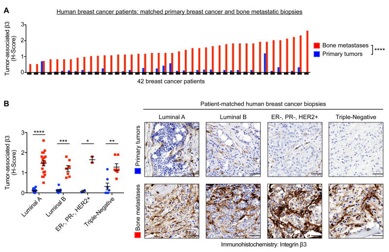
Figure 2.
Immunohistochemistry for β3 on patient-matched primary breast cancer and bone metastatic biopsies. (A) Semi-quantitatively analysis of the extent and intensity of tumor-associated β3 expression, using the histoscore (H-score) system, n=42 matched-pairs, two-tailed Wilcoxon signed-rank test, **** P<0.0001. (B) Subdivision of Fig. 2A by molecular subtype (see Materials and Methods for details). Representative images of patient-matched primary tumors and bone metastases (right). Scale=50 μm. Two-tailed paired t-test, **** P<0.0001, *** P<0.001, ** P<0.01, * P<0.05. Data presented as mean±SEM.

