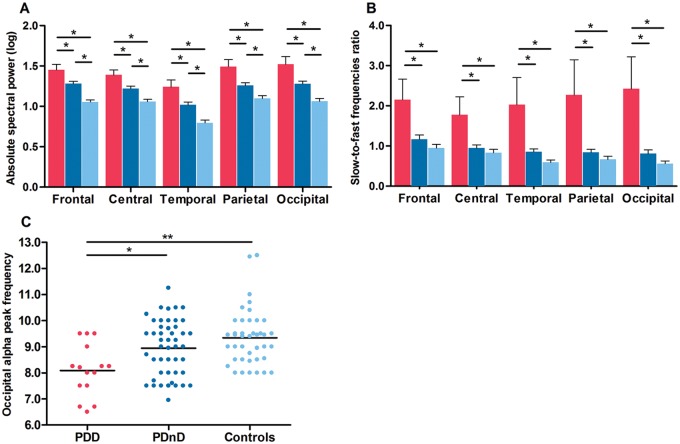
Figure 2.
PDD patients showed higher wake EEG slowing, paralleled by a lower occipital alpha peak frequency. ( A ) Delta absolute spectral power and slow-to-fast frequencies ratio ( B ) for each derivation in PDD patients (red), PDnD patients (dark blue), and controls (light blue). Post hoc analyses: * P < 0.05. Results are expressed as mean (± standard error of the mean). ( C ) Scatter plot distribution of all subjects showing lower mean alpha peak frequency at occipital electrodes (black reference line at the mean of data), * P < 0.05; ** P < 0.001.

