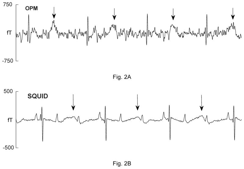
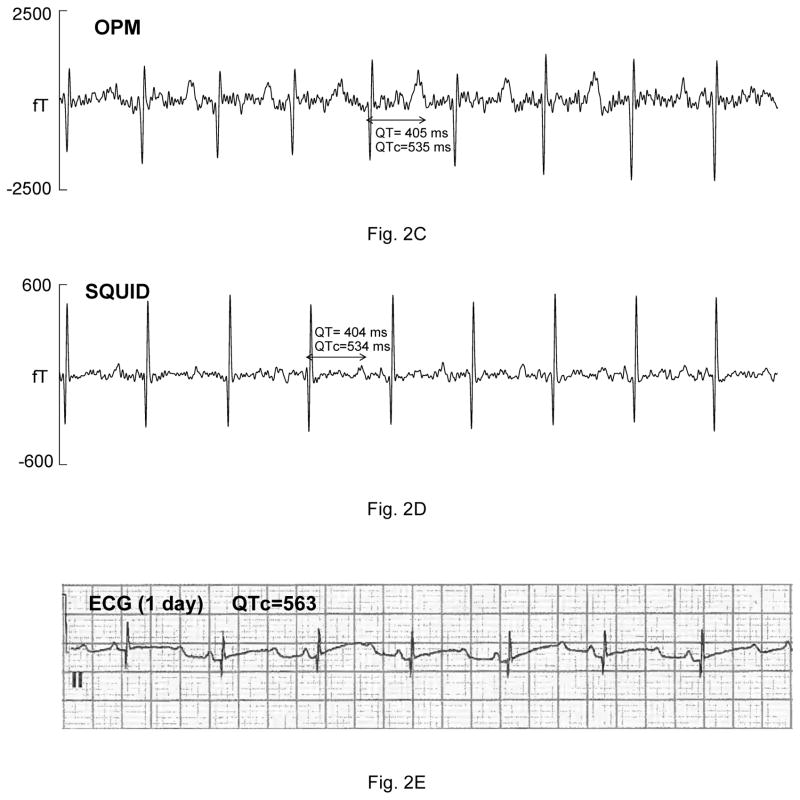
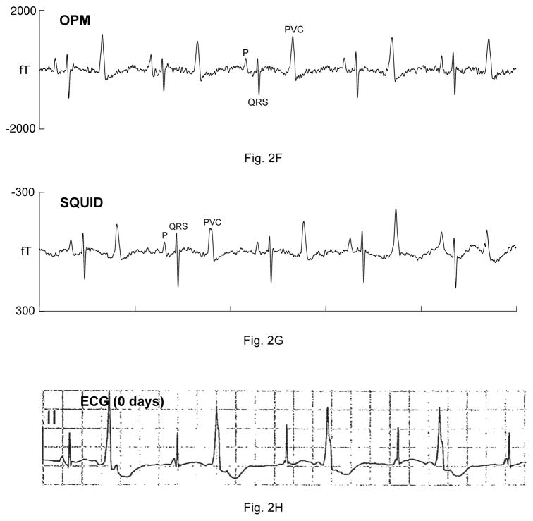
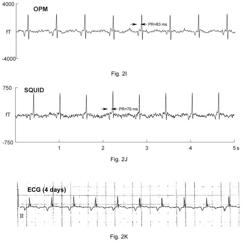
Figure 2.
Comparison of OPM and SQUID recordings from four fetuses with sustained arrhythmia. Postnatal ECGs are shown for the last three. All of the tracings are 5 seconds long. A) and B) are from a fetus at 33-4/7 weeks’ with extreme QTc prolongation (QTc> 700 ms), resulting in 3:1 AV block. The T-waves are indicated by arrows. C) and D) are from a fetus at 29 weeks’ with QTc prolongation. F) and G) are from a fetus at 33-6/7 weeks’ with a complex, irregular rhythm. As shown, the predominant rhythm was ventricular bigeminy, in which a sinus beat alternates with a premature ventricular contraction. I) and J) are from a fetus at 30-1/7 weeks’ with a low atrial rhythm, characterized by a low heart rate, inverted P-wave, and short PR interval.


