Figure 1.

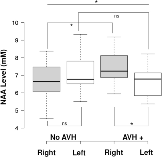
N-AcetylAspartate (NAA) levels measured by 1H Magnetic Resonance Spectroscopy in the left and right Dorsolateral Prefrontal Cortex (DLPFC) in patients with schizophrenia with (AVH+, n = 15) and without (no-AVH, n = 12) auditory verbal hallucinations (AVH). NAA levels were higher in the right DLPFC in AVH+ patients than in non-AVH patients and in the left DLPFC (F(1,25) = 5.808; partial η² = 0.189; p = 0.024). Center lines show the medians; box limits indicate the 25th and 75th percentiles; whiskers extend 1.5 times the interquartile range from the 25th and 75th percentiles.
