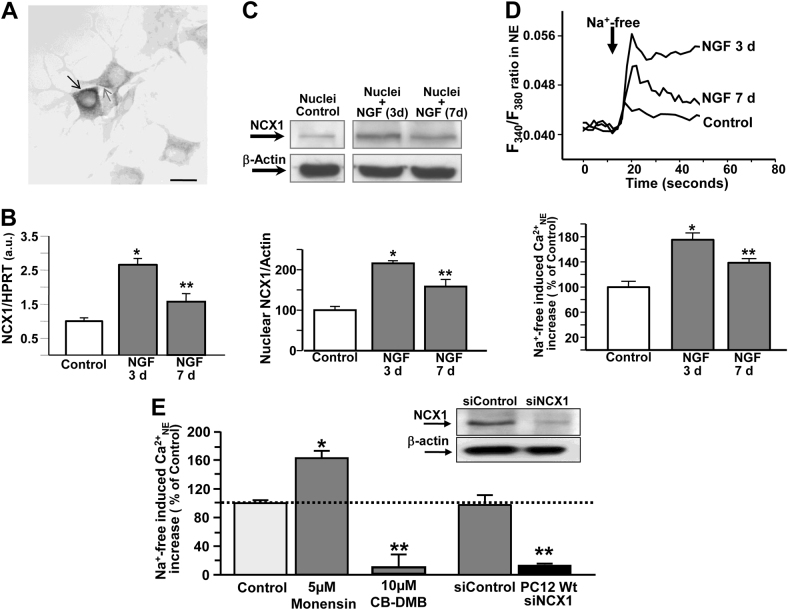
Fig. 2. nuNCX1 expression and activity in nuclei from PC12 cells during differentiation with NGF.
a Immunolocalization of NCX1 in whole cell obtained by 3,3′-diaminobenzidine in PC12 cells treated with NGF for 3 days. Scale bars: 50 µm (a), 20 µm (b). b Representative qRT-PCR of ncx1 transcript expression in control cells and in PC12 cells after 3 and 7 days exposure to NGF. c Representative Western blot and quantification of nuNCX1 protein expression in isolated nuclei from PC12 cells in control conditions and after 3 and 7 days exposure to NGF. For b and c: all the experiments were repeated at least three times on different nuclear preparations; *p < 0.05 vs. undifferentiated control, **p < 0.05 vs. undifferentiated control and 3 days NGF. d Top: Representative traces of the effect on [Ca2+]n of Na+-free perfusion in Fura-2-loaded nuclei isolated from undifferentiated PC12 cells (control), and PC12 cells exposed to 3 days or 7 days to NGF. Bottom: quantification of the effect of d. *p < 0.001 vs. undifferentiated control, **p < 0.05 vs. undifferentiated control and 3 days NGF. e Quantification of the effect on [Ca2+]n of Na+-free in isolated nuclei obtained from NGF-differentiated PC12 cells pre-treated with: monensin (10 min), CB-DMB (20 min), and siControl or siNCX1 (at DIV 5). *p < 0.001 vs. undifferentiated control, **p < 0.001 vs. all groups. Inset: Representative Western blot depicts the effect of siControl and siNCX1 on nuNCX1 protein expression in isolated nuclei from NGF-differentiated PC12 cells

