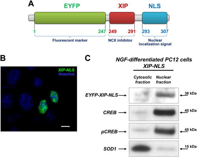
Fig. 4. Subcellular localization of the chimeric protein XIP-NLS.
a Scheme of XIP-NLS structure. b Immunolocalization of XIP-NLS (green, 400 ng/ml) detected 48 h after transfection. Hoechst dye (blue) was used to mark nuclei. Under these conditions, EYFP-positive nuclei were at least 25 ± 2% of total Hoechst-positive nuclei. c Western blot of EYFP, CREB, pCREB, and SOD1 in cytosolic and nuclear fractions obtained from NGF-differentiated PC12 cells transfected with XIP-NLS (400 ng/ml). Fractions were prepared 48 h after transfection

