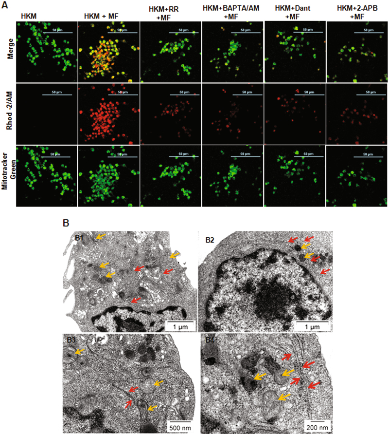
Fig. 2. Close apposition of ER and mitochondria leads to mitochondrial-Ca2+ overload in M. fortuitum infected HKM.
a HKM pre-treated with or without indicated inhibitors were infected with M. fortuitum and mitochondrial-Ca2+ uptake studied 6 h p.i. by Rhod-2/AM and Mitotracker green marker. The images are representative of three independent experiments and observed under confocal microscope ( × 40). b Transmission electron microscopy of uninfected HKM (B1), M. fortuitum infected HKM at 6 h p.i. (B2) and 24 h p.i. (B3, B4). The images are representative of three independent experiments. HKM, control headkidney macrophage; HKM + MF, HKM infected with M. fortuitum; HKM + RR + MF, HKM + BAPTA/AM + MF, HKM + Dant + MF, HKM + 2-APB + MF, HKM pre-treated with RR, BAPTA/AM, Dant and 2-APB respectively and infected with M. fortuitum. Yellow arrow,mitochondrion; Red arrow, ER

