Figure 5.
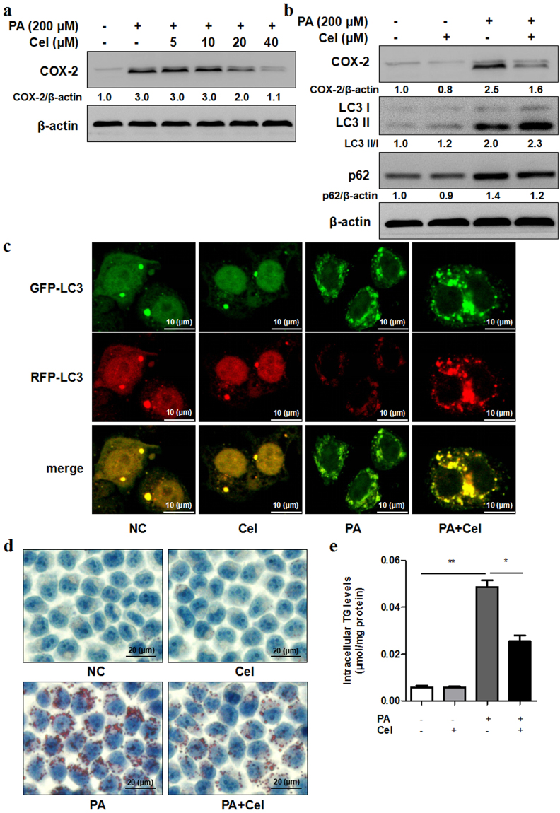
Celecoxib alleviates steatosis by restoring autophagic flux in vitro. (a) L02 cells exposed to PA (200 μM) with different concentrations of celecoxib (Cel, 5–40 μM) for 24 h. Celecoxib decreased protein expression of COX-2 compared with control as indicated by western blot. (b) L02 cells were treated with PA (200 μM) and celecoxib (20 μM) for 24 h. Celecoxib combined with PA treatment induced higher protein expression of LC3 II/I and lower protein expression of p62 compared to PA treatment. The value under the band is the ratio of the blot and normalized to control. (c) RFP-GFP-LC3 double fluorescence lentivirus tranfected L02 cells were treated with PA (200 μM) and celecoxib (20 μM) for 24 h. Representative confocal images of GFP-LC3 puncta (green, autophagosomes) and RFP-LC3 puncta (red, autolysosomes), as well as overlay images were shown (1000X). (d,e) L02 cells exposed to PA (200 μM) with celecoxib (20 μM) for 24 h. Intracellular lipid droplets accumulation and TG levels were determined by Oil Red O staining (400X) or TG assay respectively (*P < 0.05, **P < 0.01).

