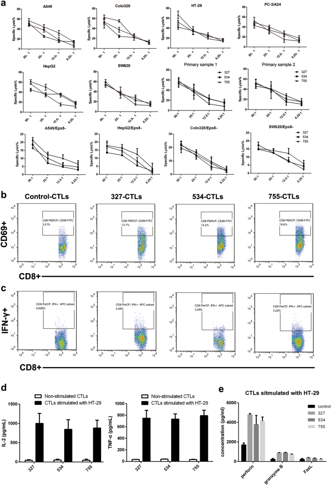
Fig. 2. Peptide-specific CTLs exhibit CTL-activating effects on various cancer cells.
a CTLs were subjected to A549, Colo320, HepG2, SW620, PC-3/A24, HT-29 cells and patient-derived tumor cells (HLA-A24+, Eps8+), as well as Eps8-silenced cells (HLA-A24+, Eps8-) at the indicated effector/target ratios (50:1, 25:1, 12.5:1, 6.25:1) by means of a standard LDH release assay. b The CD69 expression on the cell surface was evaluated after peptide stimulation by flow cytometry. c Representative flow cytometric analysis demonstrates increased IFN-γ by CTL in response to HLA-A24+ cancer cells (HT-29). CTLs showed a significantly higher level of IFN-γ secretion (*P < 0.05) in response to Eps8+ and HLA-A2+ cancer cells as compared to that in response to control CTLs cultured in media alone. d, e IL-2, TNF-α, granzyme B, perforin and FasL secretion levels by CTLs were measured by ELISAs in the cultured supernatants collected 24 h following stimulation with tumor cell lines

