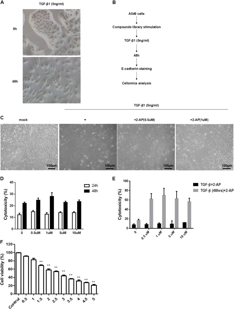
Fig. 1. Identification of compounds that regulate the TGF-β1-induced EMT.
a A549 cells were incubated with TGF-β1 (5 ng/mL) for 48 h, and the mean fluorescence intensity was determined by cellomics analysis. **P < 0.01. b Compounds were screened according to the procedure shown; the mean fluorescence intensity was detected by cellomics analysis. c A549 cells were treated with 2-AP (0.5 or 1 µM) and TGF-β1 (5 ng/mL), and morphologic changes were observed. d The effects of 2-AP on cell toxicity were determined by the LDH assay. A549 cells were treated with 2-AP at concentrations of 0 (DMSO was used as a control), 0.5, 1, 5, and 10 µM for 24 or 48 h. e The effects of 2-AP on cell toxicity in the context of TGF-β1 stimulation were determined with the LDH assay. A549 cells were treated with 2-AP at concentrations of 0 (DMSO was used as a control), 0.5, 1, 5, and 10 µM for 48 h; 2-AP was delivered either simultaneously with TGF-β1 (black bars) or 48 h after TGF-β1 stimulation (gray bars). f The effects of 2-AP on cell viability were determined by the MTT assay in A549 cells stimulated with 5 ng/mL of TGF-β1. Cells also were treated with 2-AP at concentrations of 0 (DMSO was used as a control), 0.5, 1, 1.5, 2, 2.5, 3, 3.5, 4, 4.5, or 5 µM for 48 h. **P < 0.01. Values are represented as the mean ± SD of three independent experiments performed in triplicate

