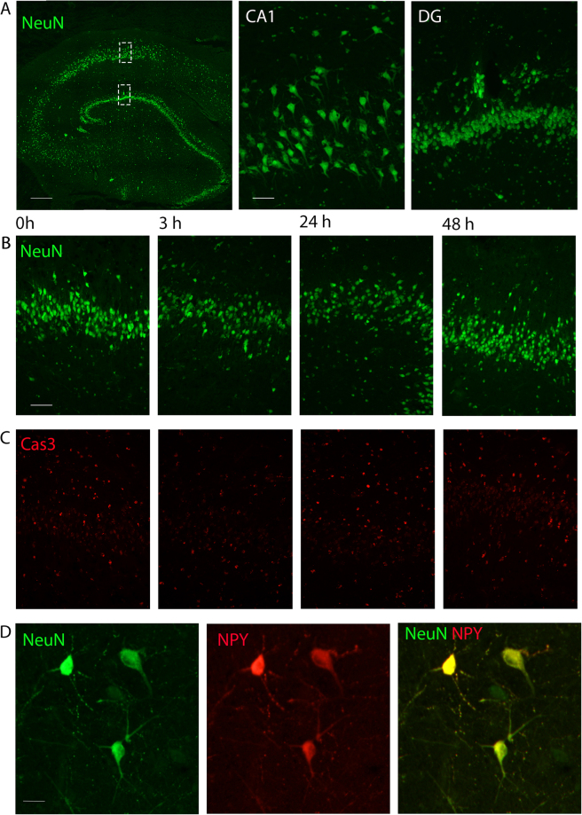
Figure 3.
Human hippocampus shows no difference in the number of neurons, expression of apoptotic marker or NPY-interneuron number in interface incubation. (A) Neuronal nuclear marker, NeuN in green (scalebar 500 μm), outlining cell layers magnified to the right for CA1 and dentate gyrus (scalebar 100 μm). (B) No differences were found in number of NeuN-positive neurons in the dentate granular layer over the time studied (green, scalebar 100 μm, Patient 16), (C) nor in the number of neurons positive for the apoptotic marker Cas3 (red). (D) Example of NPY-expressing interneurons positive for NeuN (green) and NPY (red) found in the hilar region of dentate gyrus from 48 h time-point. Scalebar 20 μm.

