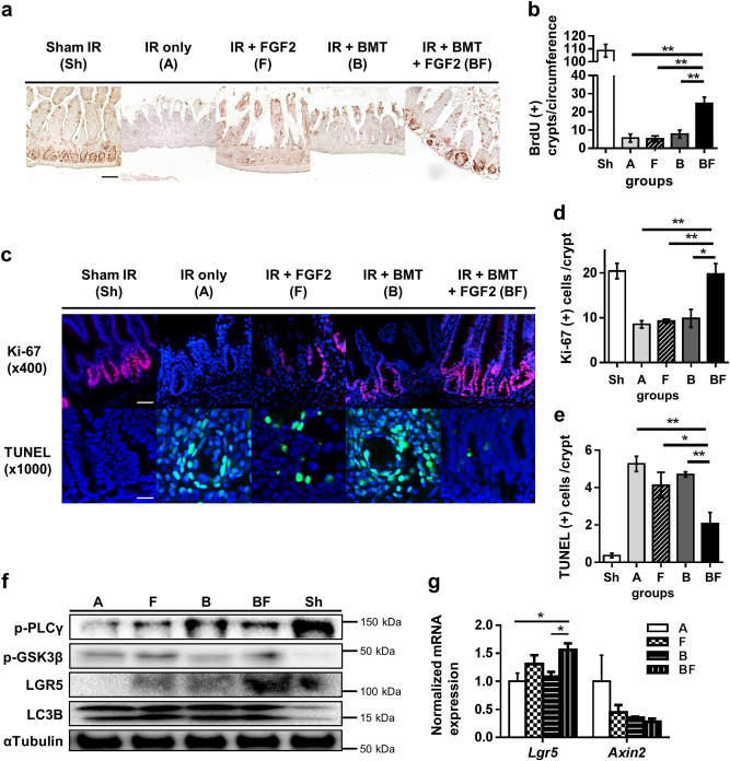
Fig. 4. Effects of FGF2 treatment combined with BMT on intestinal crypt proliferation, apoptosis, and LGR5 expression.
a BrdU incorporation measured by immunohistochemistry (magnification ×200, scale bar 100 μm) at day 5 post-irradiation. b Quantification of BrdU-positive crypts per circumference. At least five circumferences per mouse were counted; n = 3 in each group. c Ki-67 and TUNEL immunofluorescence staining (Ki-67: magnification ×400, scale bar 50 μm; TUNEL: magnification ×1000, scale bar 20 μm). d, e Quantification of Ki67- and TUNEL-positive cells per crypt at day 5 post-irradiation. At least 10 well-oriented crypts per mouse were counted; n = 3 in each group. f Intestinal tissue lysates were examined by Western blot at day 3 post-irradiation. α-tubulin was used as the loading control and LC3B was used as the positive control for radiation damage (autophagy). Data are representative results from at least two independent experiments. g Lgr5 and Axin2 expression levels at day 3 post-irradiation; n = 3 in each group. Values are mean ± SEM; *p < 0.05, **p < 0.01

