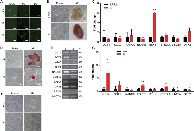
Fig. 6. Maintenance of porcine iPS cell lines in 3i medium.
a Fluorescence analysis of OCT4 promoter (PE/DE), OCT4 proximal enhancer (PE), and distal enhancer (DE) in DOX-iPSCs with different media. b Morphology and alkaline phosphatase (AP) staining of LFB2i-piPSCs grown in LFB2i and 3i media. c Quantitative RT-PCR analysis of pluripotent genes from LFB2i-piPSCs in LFB2i and 3i media. d Morphology and AP staining of iPF4-2 cells grown in 2i and 3i media. e RT-PCR analysis of pluripotent genes from iPF4-2 cells grown in 2i and 3i media. f Morphology and AP staining of pPSCs grown in MXV and 3i media. g Quantitative RT-PCR analysis of pluripotent genes from pPSCs in MXV and 3i media. Scale bar, 100 μm. Data indicate mean ± SD. *P < 0.05, **P < 0.01, n = 3

