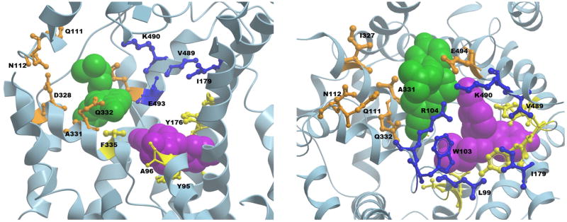
Figure 5.
Side (left) and top (right) views of the human SERT crystal structure bound with two escitalopram molecules (PDB ID 5I73), with a few protein backbone structures truncated for the ease of visibility. One escitalopram molecule (purple) is bound to the primary, high-affinity S1 binding site with residues shown in yellow. The residues of the secondary allosteric site SERT/A1 in the extracellular vestibule are depicted in blue. The SERT/A2 site residues are shown in orange. The second escitalopram molecule of the crystal structure bound in the extracellular vestibule is shown in green.

