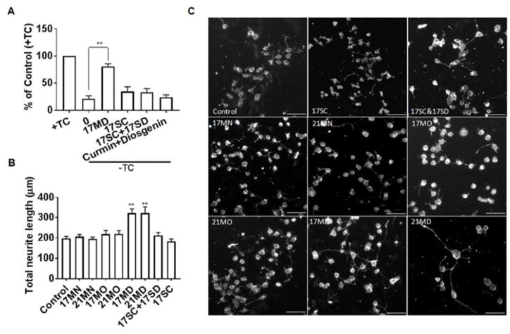
Figure 3. Protective activities in MC65 cells and neuritic outgrowth stimulating effects in N2a cells by tested compounds.
(A) MC65 cells were treated with indicated compounds or the combination of indicated compounds (0.3 μM) under –TC conditions for 72 h. Then cell viability was measured by MTT assay. Data were presented as mean (n=3) ± SEM (**p < 0.01 compared to -TC). (B) Neuronal N2a cells were seeded in 24-well plates and treated with indicated compounds (0.3 μM) for 48 h. Cells were stained with the Neurite Outgrowth Staining kit (Life Technologies) and images were recorded by Zeiss Axiovert 200M fluorescence microscopy using 10× objective and average neurite length was analyzed using Image J program. Data were presented as mean (n=3) ± SEM (**p < 0.01 compared to control). (C) Representative images from three independent experiments. Scale bar: 100 μm.

