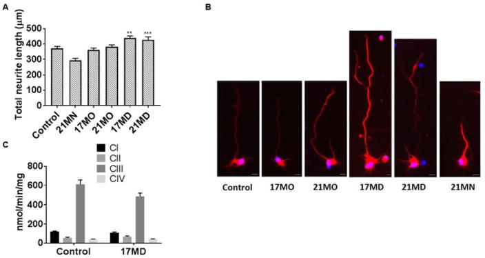
Figure 4. Effects on neuritic outgrowth of mouse primary cortical neurons and the enzymatic activity of ETC complexes of mouse brain mitochondria.
(A) Cortical neurons from E17 embryos were treated with DMSO or compounds for 4 days. Neurons were immunostained with the neuronal marker beta III tubulin (red) and nuclei were detected by use of DAPI (blue). Quantification of the average length of the total neurites was quantified by Image J analysis. Data were presented as mean ± SEM) (** P < 0.01, *** P < 0.001). (B) Representative images of neurons for the corresponding groups to illustrate the length of neurite are shown in (A). Scale bar: 20 μm. (C) Detergent solubilized mouse brain mitochondria were treated with 17MD (3.3 μM) and the respiratory complex activities were determined using spectrophotometer. No difference in complex I, II, III, and IV was observed between control and 17MD treated mitochondria. Data were presented as mean ± SEM (n=4).

