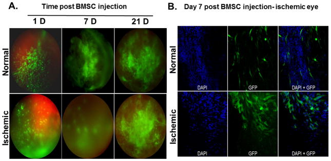
Figure 4.
Ischemic and normal eyes were injected with GFP-BMSC 24 h post ischemia. In vivo imaging of the eyes was performed at 1, 7 and 21 days’ post injection (A). The upper and lower panels exhibit representative images of normal and ischemic eyes injected with BMSCs. (B) Representative flat mount images of BMSCs injected retina illustrate the penetration and incorporation of significantly increased number of GFP-BMSCs into the ischemic retinal tissue 7 days’ post injection compared to normal retina. DAPI was used to stain nuclei.

