Figure 5.
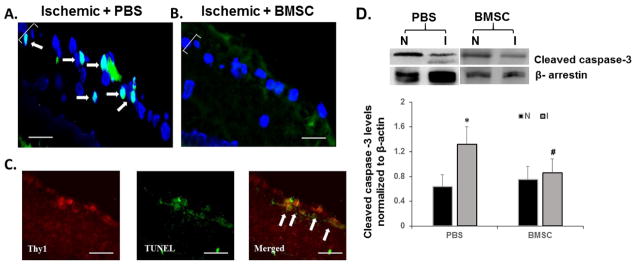
Fluorescent TUNEL staining for evaluation of apoptosis in 10-μm thick retinal cryosections: BMSC injection into the retina significantly reduced apoptotic cell death in the ischemic retinae 24 h post injection (B) as compared to PBS injected ischemic control (A). White arrows denote TUNEL positive cells (green) co-localized with DAPI (blue). Brackets denote the retinal ganglion cell layer. Magnification = 40X. Scale bar = 15 μm. (C) Retinal ganglion cells undergoing apoptosis in PBS injected control ischemic retina identified by co-localization of TUNEL and Thy1 (retinal ganglion cell marker, red). Magnification = 40X. Scale bars = 20 μm. (D) Representative Western blot images for cleaved caspase-3 in retinal lysates obtained from PBS or BMSC ischemic injected eyes and paired normal eyes, at 24 h after injection of PBS or BMSCs. Levels of cleaved caspase-3 were normalized to β-arrestin. Data are shown as mean ± SEM. * indicates P < 0.05 for ischemic compared to the paired normal retina within a group (PBS or BMSC); # indicates P < 0.05 between the ischemic retinae of PBS and BMSC. N = normal, non-ischemic retinae, and I = ischemic retinae. N = 5 per group.

