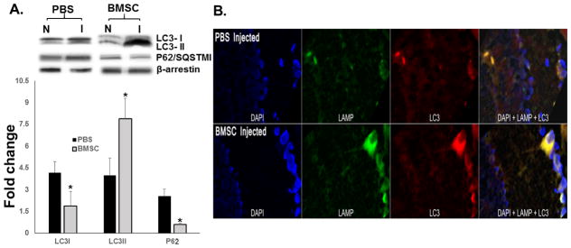
Figure 7.
(A) Western blotting for LC3-I, LC3-II, and p62 as markers of autophagy. The results are expressed as fold change from normal to ischemic within each group, with protein levels normalized to β-arrestin. N = 5 per group. Data are shown as mean ± SEM. There were significant decreases in LC3-I and in p62, and significant increases in LC3-II in the BMSC group. * indicates P < 0.05 compared to the paired normal retina within a group (PBS or BMSC); # indicates P < 0.05 between the ischemic retinae of PBS and BMSC. (B) Retinal cryosections stained for DAPI (blue), LAMP (red), and antibody specific for LC3-II (green), in PBS injected (top), and BMSC injected (bottom) ischemic retinae. The images demonstrate that LC3-II and LAMP co-localize in cytoplasm, indicating their localization into autophagosomes, confirming the presence of autophagy. See Methods for more details.

