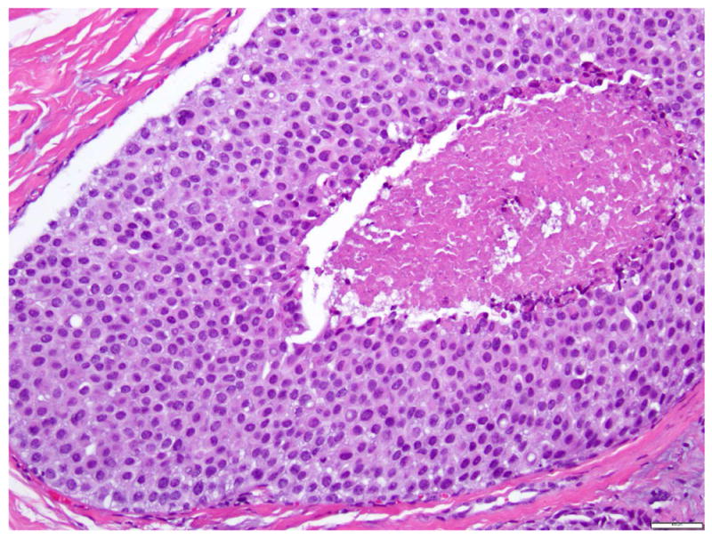
Fig 10. Lobular carcinoma in situ with central necrosis.

This proliferation of cells morphologically indistinguishable from those of classic LCIS, is associated with massive acinar expansion (50 or more cells across the diameter of an expanded acinus) and central necrosis. Magnification 200x.
