
Fig 13. Pleomorphic lobular carcinoma in situ, apocrine type.
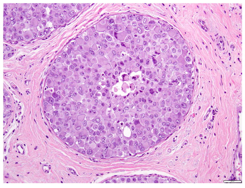
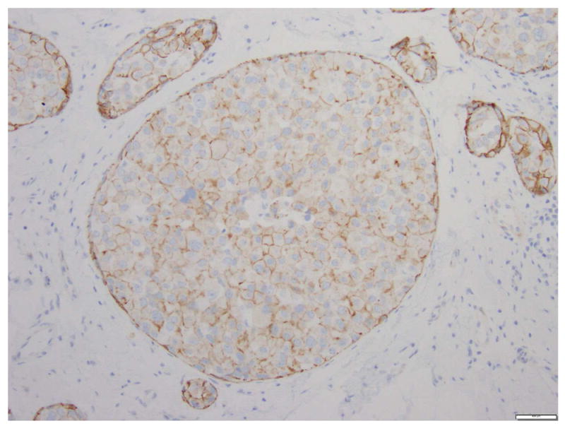
a. H&E stain. b. Immunohistochemical stain for E-cadherin. The cells are dyshesive, with abundant eosinophilic granular cytoplasm, large nuclei, and prominent nucleoli. Instead of complete loss of E-cadherin expression, the cells composing PLCIS in this case show focal incomplete, attenuated, and granular membranous staining for E-cadherin. Magnification 200x.