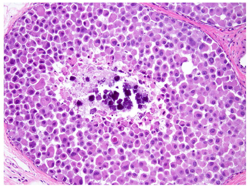
Fig 8. Lobular carcinoma in situ, pleomorphic type.

Dyshesive proliferation of round to oval cells with abundant cytoplasm, large eccentric nuclei with irregular nuclear membrane, coarse chromatin, and prominent nucleoli. Foci of necrosis with calcifications are common. Magnification 200x.
