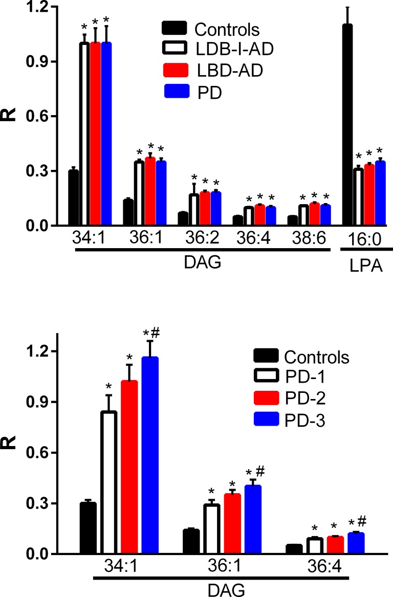
Fig 1. Frontal cortex levels of diacylglycerols (DAG) and lysophosphatidic acid 16:0 (LPA) in control, Lewy body disease with intermediate Alzheimer’s disease (LBD-I-AD), Lewy body disease with Alzheimer’s disease (LBD-AD), and Parkinson’s disease (PD) tissues.
R = ratio of the peak area of the endogenous lipid to the peak area of the internal standard (mean ±SEM). Analysis of PD subgroups demonstrated that DAGs were augmented in the cortex of PD subjects with no neocortical pathology (PD-1, N = 5), subjects with sparse neocortical neuritic plaques (PD-2, N = 5), and subjects with moderate to frequent neocortical neuritic plaques (PD-3, N = 5). *, p < 0.01 vs. controls; #, p < 0.05 for PD-2 vs. PD3.

