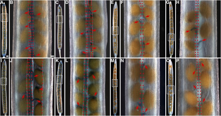
Figure 2.
GUS staining patterns in siliques of the GUS-promoter and GUS-protein fusion lines of the CEL6 and MAN7 genes in the wild-type, ind-7, and alc-3 backgrounds. A and B, Siliques of a pCEL6:GUS line. C and D, Silique of a pCEL6:CEL6-GUS line. E and F, Silique of a pCEL6:GUS line in the ind-7 mutant background. G and H, Silique of a pCEL6:GUS line in the alc-3 mutant background. I and J, Silique of a pMAN7:GUS line. K and L, Silique of a pMAN7:MAN7-GUS line. M and N, Silique of a pMAN7:GUS line in the ind-7 mutant background. O and P, Silique of a pMAN7:GUS line in the alc-3 mutant background. B, D, F, H, J, L, N, and P, Higher magnification images of the rectangular areas in the images immediately to the left, respectively. Red arrows indicate funiculi. In dehisced siliques (B, D, and J), vertically aligned red dots delineate the replum (right two dotted lines in B or two central dotted lines in D and J) and mark the freed valve margins. In nondehisced siliques (F, H, L, N, and P), the dotted lines mark the junction areas between the valves and the replum. Bar in A = 500 μm for A, C, E, G, I, K, M, and O and bar in B = 100 μm for B, D, F, H, J, L, N, and P.

