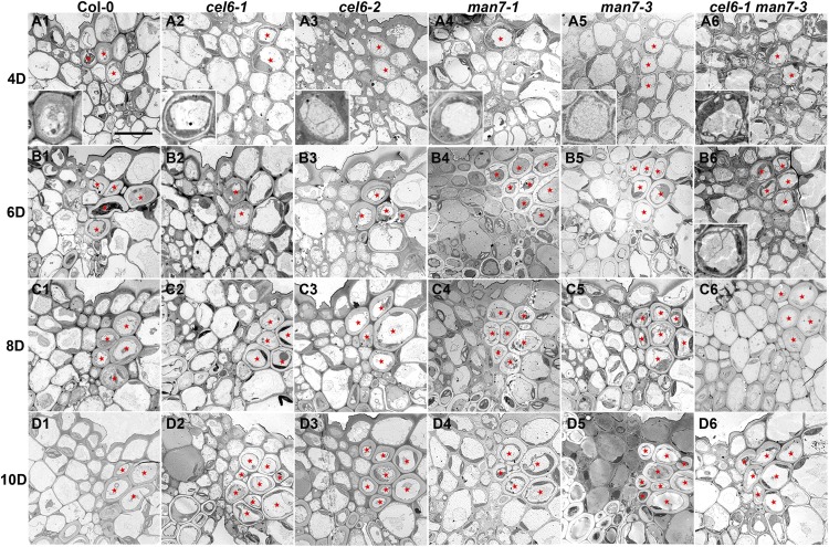
Figure 4.
Transmission electron microscopy images of transverse sections of stage-17 siliques of Col-0 and the cel6 and man7 mutants. A1 to D1, Col-0. A2 to D2, The cel6-1 mutant. A3 to D3, The cel6-2 mutant. A4 to D4, The man7-1 mutant. A5 to D5, The man7-3 mutant. A6 to D6, The cel6-1 man7-3 double mutant. A1 to A6, Siliques at stage 4D (4 d after anthesis). B1 to B6, Siliques at stage 6D. C1 to C6, Siliques at stage 8D. D1 to D6, Siliques at stage 10D. Red stars in Col-0 indicate lignified cells, and in the mutants either cells presumably destined to be lignified or lignified cells, in the dehiscence zone. Insets in A1 to A6 and B6 show enlargements of such cells in the corresponding images. Bar = 10 μm for all images (excluding the insets).

