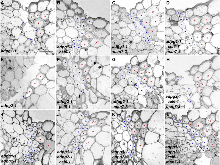
Figure 8.
Transmission electron microscopy images of transverse sections of stage 18B siliques in single, double, triple, and quadruple mutants. A, The adpg1-1 mutant. B, The cel6-1 adpg1-1 double mutant. C, The man7-3 adpg1-1 double mutant. D, The cel6-1 man7-3 adpg1-1 triple mutant. E, The adpg2-1 mutant. F, The cel6-1 adpg2-1 double mutant. G, The man7-3 adpg2-1 double mutant. H, The cel6-1 man7-3 adpg2-1 triple mutant. I, The adpg1-1 adpg2-1 double mutant. J, The cel6-1 adpg1-1 adpg2-1 triple mutant. K, The man7-3 adpg1-1 adpg2-1 triple mutant. L, The cel6-1 man7-3 adpg1-1 adpg2-1 quadruple mutant. Red stars indicate lignified cells in the dehiscence zone. Blue triangles indicate presumed broken or intact separation layer cells. Bar = 10 μm for all images.

