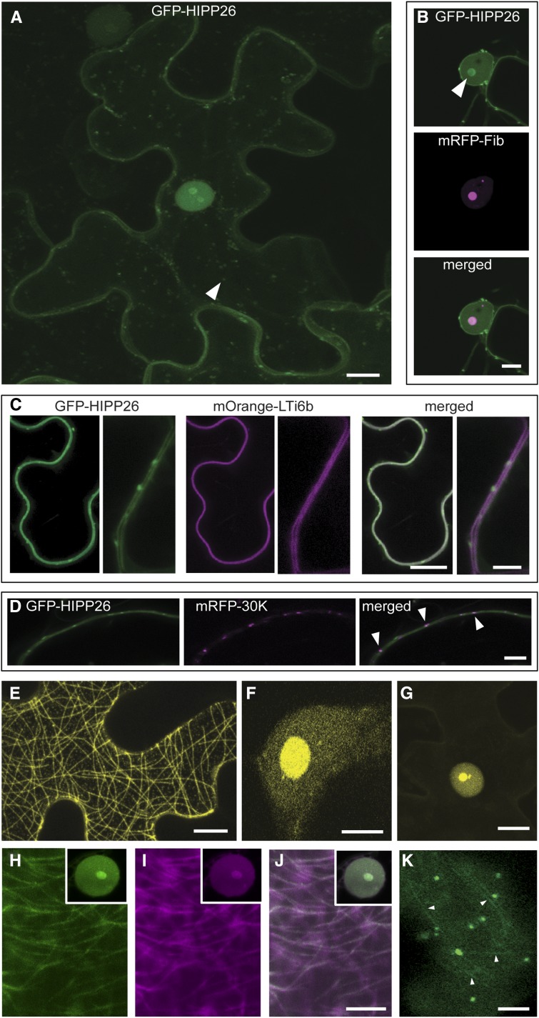
Figure 3.
Subcellular localizations of HIPP26 and the HIPP26-TGB1 complex. A, The GFP-HIPP26 fusion protein localized to the plasma membrane and a population of small (∼2 µm diameter), motile vesicles (arrowhead). A small amount of cytosolic labeling and faint transvacuolar strands were seen in some cells. GFP-HIPP26 also was present in the nucleus, both in the nucleoplasm and the nucleolus, and to levels greater than would be expected as a result of passive diffusion from the low levels in the cytosol. Therefore, HIPP26 was targeted to the nucleus. B and C, Coexpression with an mRFP-fibrillarin (a nucleolar marker) showed precise colocalization of GFP-HIPP26 in the nucleolus (B; indicated by the arrowhead), while coexpression and colocalization with mOrange-LTi6b (a plasma membrane marker) showed that HIPP26 also was targeted to the plasma membrane (C). D, In addition to the motile vesicles that were seen in the cytoplasm of the cell periphery (compare with A and Supplemental Movie S1), small nonmotile punctae also were observed at the cell periphery. Colocalization of these structures with an mRFP-tagged TMV 30K protein (a PD marker) showed that HIPP26 also was associated with a population of PD (indicated by arrowheads). E and F, BiFC analysis of PMTV TGB1 and NbHIPP26 in N. benthamiana epidermal cells showed coexpression of Y-HIPP26 and TGB1-FP and reconstitution of yellow fluorescence localized to microtubules (E), to the nucleoplasm, and strong accumulation in the nucleolus (F). G, Coexpression of Y-HIPP26 and ∆84TGB1-FP showed reconstitution of yellow fluorescence localized to the nucleoplasm and strong accumulation in the nucleolus with no microtubule labeling. No complementation was obtained in control experiments, where Y-HIPP26 or TGB1-FP was coexpressed with the complementary empty split YFP plasmid (no fluorescence was visible, so no images are shown). H to J, Colocalization of GFP-HIPP26 and mRFP-TGB1 (H, GFP; I, RFP; and J, merged) showed that both proteins localized to the nucleoplasm, nucleolus, and microtubules, as expected from previously published data and the BiFC results. K, Expression of GFP-HIPP26 in a PMTV-infected cell showed that GFP fluorescence accumulated on microtubules (indicated by arrowheads). Bars = = 10 µm in A, C (at left), D, E, G, and J (used for H–J) and 5 µm in B, C (at right), F, and K.

