Figure 2.
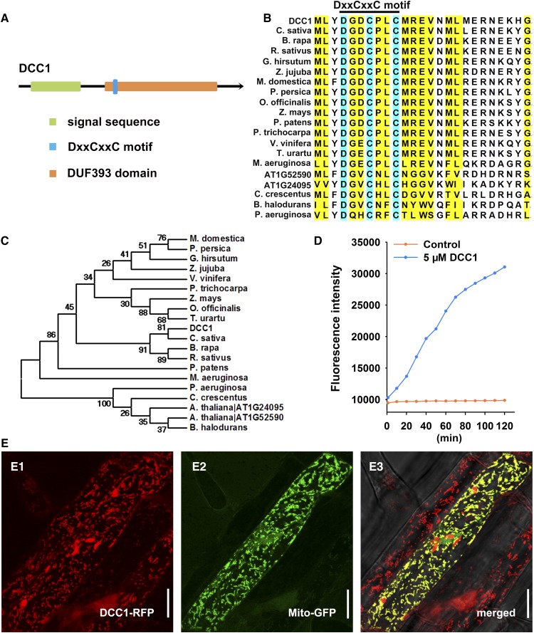
DCC1 is a functional Trx localized in mitochondria. A, Model of the DCC1 protein with a mitochondria signal sequence predicted in the UniProt database and a conserved DxxCxxC motif in a function-unknown DUF393 domain analyzed in the Pfam database. B, Alignment of the amino acid sequences of different DCC family proteins. The DxxCxxC motif is a conserved signature sequence for DCC family proteins in various species. The National Center for Biotechnology Information accession numbers of proteins in different species are presented in “Materials and Methods.” C, Phylogenetic analyses of DCC1 with its homologs in various species. DCC1 shares high homology with several proteins in species such as C. sativa, B. rapa, and R. sativus. D, Insulin reduction by recombinant DCC1 proteins. Purified DCC1 proteins were subjected to a reduction assay by using FiTC-insulin as the substrate, which displayed higher fluorescence after disulfide reduction. The assay mixture lacking recombinant DCC1 proteins served as the control. Fluorescence intensity was recorded at 515- to 525-nm emission after 480- to 495-nm excitation for 120 min in a fluorescent plate reader at room temperature. E, Subcellular localization of DCC1. MT-GK is a well-established marker line specially expressing Mito-GFP. The construct 35S:DCC1-RFP was transformed into MT-GK. The T1 transgenetic line roots were excised for imaging by a confocal microscope. The DCC1-RFP signal (E1) was observed at 505- to 550-nm emission after 561-nm excitation, whereas the Mito-GFP signal (E2) was observed at 570- to 620-nm emission after 488-nm excitation. The merged signals of GFP and RFP showed yellow color (E3), indicating that DCC1 is localized in the mitochondria. Bars = 20 µm.

