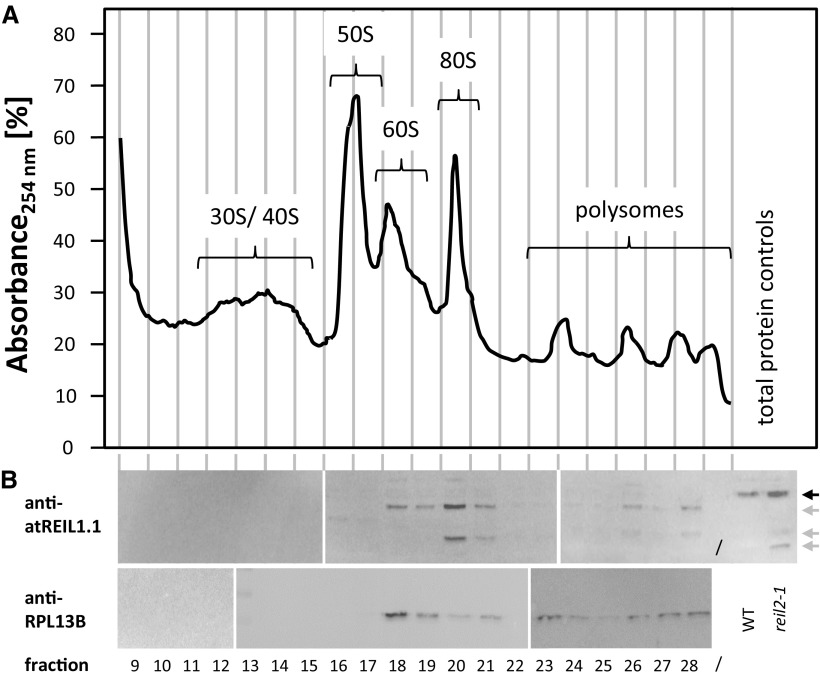
Figure 6.
Western-blot analysis of Col-0 ribosome fractions. Ribosome protein fractions from the Col-0 wild type were compared with total protein preparations from Col-0 (WT) and reil2.1. All preparations were from rosette plants of stage ∼1.10 that were cultivated at 20°/18°C (day/night). A, Absorbance profile of the Suc density sedimentation gradient analysis at wavelength λ = 254 (A254) after blank gradient subtraction. Vertical lines indicate the approximate positions of the collected protein fractions. B, Western-blot analyses of the indicated fractions using anti-atREIL1.1 (top) and anti-RPL13B (bottom) antibodies. Note that anti-atREIL1.1 was directed against a variable region at the REIL1 C terminus. This antibody detects REIL1 in total protein extracts (black arrow) and cleavage products (gray arrows) in protein preparations after Suc density gradient centrifugation. Cleavage products also are detectable at low abundance in total protein from the reil2.1 mutant. The western-blot analysis was performed by three parallel-processed blots of fractions from the sedimentation gradient shown in A. The anti-RPL13B analyses were of an independent sedimentation gradient. Splice sites of blots are indicated by white bars.

