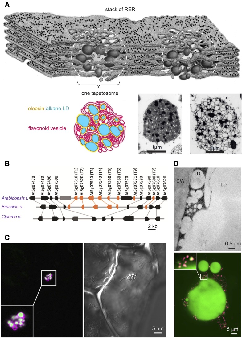
Figure 3.
Specialized LDs in restricted tissues of specific plant phylogenies. A, T oleosin-containing LDs in tapetosomes being synthesized in rough ER in the tapetum of Brassica spp. In the model, a stack of rough ER produces alkane-containing, T oleosin- and PL-coated LDs and flavonoid-possessing vesicles at many locations. At each location (marked with a dotted white circle), numerous LDs and vesicles aggregate to form a tapetosome. The bottom shows a color model of a tapetosome having numerous alkane-containing (blue) LDs enclosed with a layer of oleosin-PL (yellow) associated with flavonoid-possessing vesicles (red). At right of the color model are electron micrographs of isolated tapetosomes without (middle) and with (right) osmotic swelling; after swelling of the tapetosome, internal LDs and vesicles can be seen. (A portion of the rough ER stack was traced from a drawing in a Trends in Cell Biology poster, 1998 [Hsieh and Huang, 2004, 2005]). B, T oleosin gene cluster in Brassicaceae and its absence in Cleomaceae in syntenic chromosome segments. Coding sequences of genes are indicated with colored arrows (orange for oleosin genes and black for nonoleosin genes) in genomic DNA from Arabidopsis, Brassica oleracea, and Cleome violacea. Orthologs among the genomes are linked with gray lines. The large gray box indicates a transposon (Huang et al., 2013a). C, U oleosin-containing LD cluster in vanilla leaf epidermis. Right, Differential interference contrast microscopy image shows the cell containing one LD cluster. Left, Immuno-CLSM image of the same cell reveals the LDs and oleosin. BODIPY-stained (in green) individual LDs in the cluster are visible. Antibodies against vanilla U oleosin reacted (in magenta) with the LDs. Oleosin appears more on the periphery of individual LDs, resulting in a magenta coat enclosing a white matrix (Huang and Huang, 2016). D, M oleosin-containing small LDs and oleosin-depleted large LD in avocado mesocarp. Top, Transmission electron microscopy image showing a portion of an avocado mesocarp cell containing large (10–50 μm) and small (less than 0.5 μm) LDs adjacent to the cell wall (CW). Bottom, Immuno-CLSM images show a large LD and numerous adjacent small LDs as well as a magnified junction between a large LD and numerous small LDs. BODIPY-stained (in green) large and small LDs are visible. Antibodies against avocado M oleosin reacted (in magenta) mostly with the small LDs. Oleosin appears more on the periphery of individual small LDs, resulting in a magenta coat enclosing a yellow matrix (Huang and Huang, 2016).

