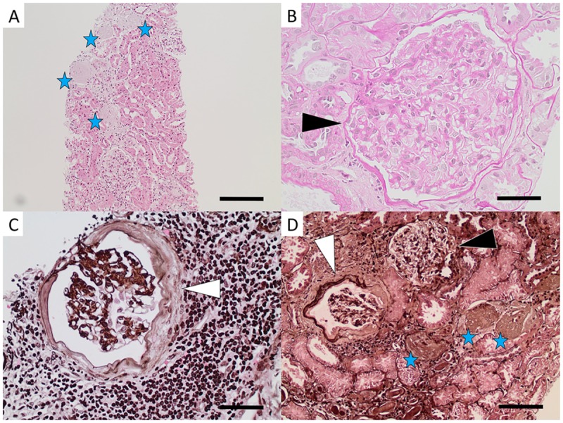
Fig 1. Light microscopy images of glomerular pathology in Sri Lankan patients with CKDu.

Global glomerulosclerosis (stars in A and D) and glomerular hypertrophy (black arrow heads in B and D) of varying degree were found in all biopsies. Signs of glomerular ischemia with thickening of Bowman’s capsule (white arrow heads in C and D) and/or wrinkling of the capillaries was seen in seven patients. [Fig A: hematoxylin-eosin from Patient 4, bar = 200μm. Fig B: periodic acid Schiff from Patient 6, bar = 50μm. Fig C: periodic acid silver methenamine from Patient 3, bar = 50μm. Fig D: periodic acid silver methenamine from Patient 6, bar = 100μm.].
