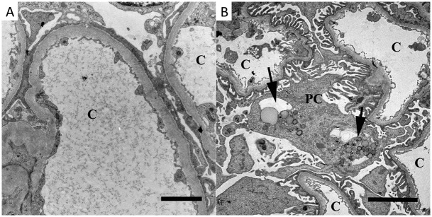
Fig 4. Transmission electron microscopy findings.
Segmental podocytic foot process effacement was observed in two patients (A, Patient 4). Podocytic cytoplasm inclusions of vacuoles or lipofuscin-like-bodies (arrows in B, Patient 6) were found in the majority of the patients. c = capillary, pc = podocyte. Bars = (A) 2μm, (B) 5 μm.

