
Figure 4. Immunohistochemical evaluation of blood brain barrier permeability.
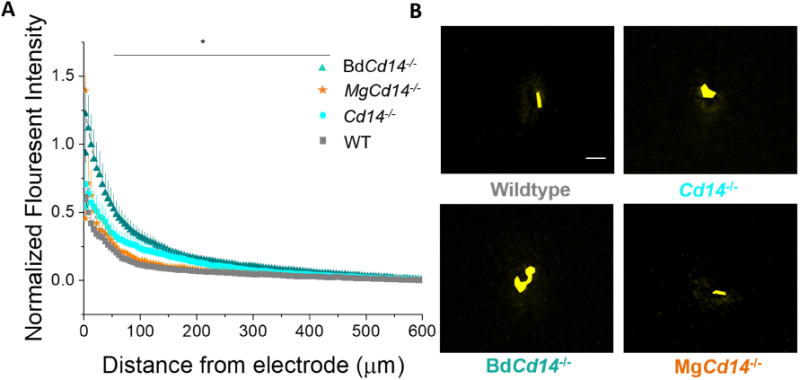
(A) Blood brain barrier permeability evaluated as IgG expression with respect to distance from the explanted microelectrode hole (μm). Significant differences between wildtype and BdCd14-/- were observed from 50-450 μm away from electrode-tissue interface, * p<0.05). (B) Representative images from tissue derived from ∼380 - 830 μm deep from surface of brain. Yellow area represents hole left by explanted probe. Scale bar: 50 μm