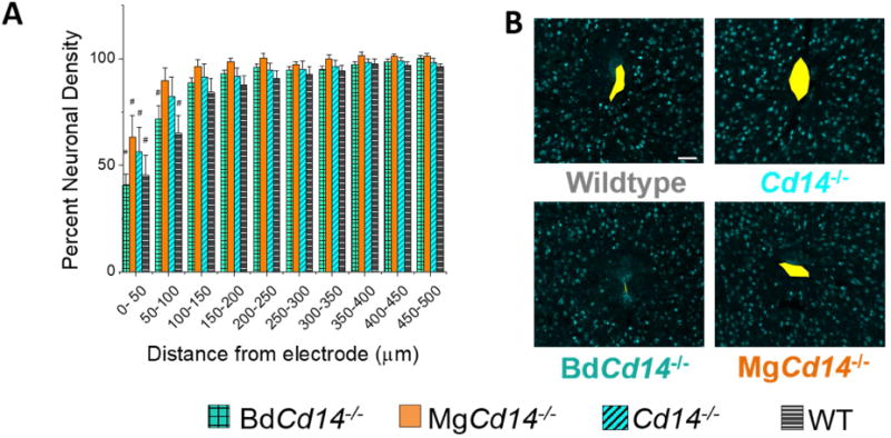
Figure 6. Immunohistochemical evaluation of neuronal density.

(A) Neuronal density evaluated as NeuN+ counts with respect to distance from the explanted microelectrode hole (μm). No significant differences were observed among experimental groups. Neuronal density is significantly different from background MgCd14-/- and wildtype between 0 and 50 μm from the microelectrode hole, and Cd14-/-and BdCd14-/- between 0 and 100 μm from the microelectrode hole, # p<0.05. (B) Representative images from tissue acquired from ∼625-825 μm deep from surface of brain. Yellow area represents hole left by explanted probe. Scale bar: 50 μm
