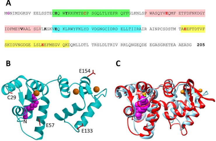
Fig 1. Primary and tertiary structure of GCAP1.
(A) Amino acid sequence of bovine GCAP1, (B) crystal structure of GCAP1 in the Ca2+-saturated state (cyan, PDB code: 2R2I), and (C) overlay of GCAP1 crystal structure (cyan) with NMR Structure of GCAP1 in the Ca2+-free/Mg2+-bound state (red, PDB code: 2NA0). EF-hand motifs in the primary sequence are shaded in color (EF1 green, EF2 red, EF3 cyan and EF4 yellow). GCAP1 residues substituted with cysteine (T29C, E57C, K67C, E133C, E154C) that are attached to a nitroxide spin-label in DEER studies are highlighted in bold and red. Key residues at the dimer interface are highlighted bold and black. N-terminal myristoyl group is colored magenta. Bound Ca2+ and Mg2+ are colored orange and green, respectively.

