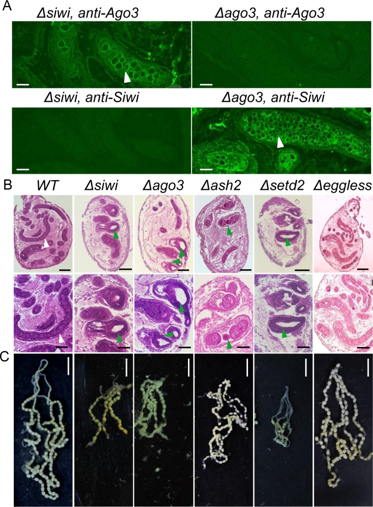
Fig 2. Arrested oogenesis in mutants.

(A) Immunohistochemistry in Δsiwi and Δago3 W stage ovaries. The white arrowheads indicate fused egg chambers and the accumulation of germline cells in the ovarioles. Scale bars represent 50 μm. (B) Paraffin-embedded sections of ovaries from WT, Δsiwi, Δago3, Δash2, Δsetd2 and Δeggless females at W stage. The lower row showed magnified images (X40). Tissues were stained with hematoxylin-eosin and photographed under a bright field. White arrowheads indicated normal ovariole structures, and green arrowheads indicated the atrophic ovarioles, which were short, vacuolated and contained fused egg chambers in the mutants. Scale bars represent 0.25 mm and 0.125 mm in the upper and lower row respectively. (C) Arrested oogenesis in Δsiwi, Δago3, Δash2 and Δsetd2 mutants. Scale bars represent 0.5 cm.
