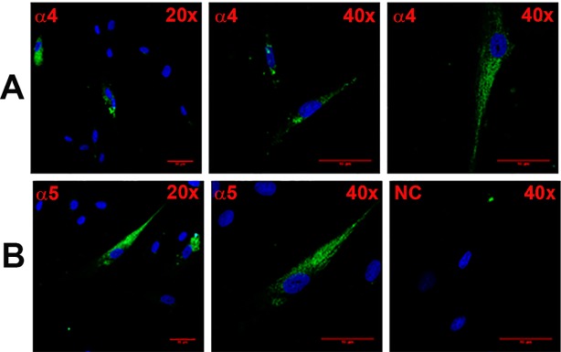
Fig 2. Immunofluorescence staining of CHRNA4 and CHRNA5 in HBO cells.
Shows immunostaining of CHRNA4 (A; α4) and CHRNA5 (B; α5) in HBO cells using 20x and 40x objectives (total 200x and 400x magnification). The panels show merged confocal images of DAPI (blue) and secondary antibody fluorescence (green). The negative control (NC) without primary antibody is also shown. The horizontal red lines represent 10 μm.

