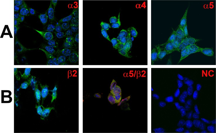
Fig 10. Immunofluorescence staining of CHRNA and CHRNB subunits in HEK293 cells.
(A) Shows immunostaining of CHRNA3 (α3), CHRNA4 (α4), and CHRNA5 (α5) in HEK293 cells. (B) Shows immunostaining of CHRNB2 (β2) in HEK293 cells. The panels show merged confocal images of DAPI (blue) and secondary antibody fluorescence (green). The negative control (NC) without primary antibody is also shown. Dual immunostaining was used to co-localize CHRNA5 (α5) with CHRNB2 (β2). (B, α5/β2) Shows immunostaining of CHRNA5 (α5; green) with CHRNB2 (β2; red). The panel shows merged confocal images of DAPI and dual fluorescence labels.

